Phone:
+48 601 333 666
Email:
office@asamed.com
Select Language
English
Polish
Inventory
Analytical Lab
Co2 Analyzer
Fume/Bio Safety Hood
Liquid Chromatograph/HPLC
Automatic lines and Sorters
Biochemistry
Analyzer
Biochemistry
Chemistry Analyzer
Blood Bank
Blood Analyzer
Blood bank
Immunology
Refrigerator Freezer
Blood Gas
Blood Analyzer
Blood Gas Analyzer
Clinical Lab
Centrifuge
Centrifuge
Rotator/Mixer/Rocker
Coagugology
Coagulation Analyzer
Coagulology
Computer Equip
Scanner
Consumables and Supplies
Disposables - General
DNA Test
Dna test
Slide Stainer
Electrolytes
Electrolyte Analyzer
Hematology
Hematology
Hematology Analyzer
Homecare/Rehab
Bath Chair
Scale
Water Purification
Immunoassay ELIZA
Immunology
Immunology ELIZA
Immunology
Immunology
Incubators
Dryer
Incubator
Mass Spectrometer
Spectrophotometer
Medical Equipment
Clinical Lab
Microbiology
Microbiology
Microscopes
Microscope
Monitors/ICU/CCU
EKG
Oximeter - Pulse
Nephelometry
DNA Related
Ophthalmology
Ophthalmoscope
Other
Other
Phmeter
Physical Therapy
Magnetic Field Therapy
Recovery Room
Semi-Automated Chemistry
Chemistry Analyzer
Serology
Serology
Spectrophotometer
Spectrophotometer
Spirometers
Spirometer
Sterilization
Test Equipment and Tools
Power Supply
Temperature/Pressure
Ultrasound
Ultrasound
Ultrasound General
Ultrasound Transducer
Vascular - Small Parts Ultrasound
Urine Analyzer
Urine Analyzer
Urine Flowcytometers
Urine Analyzer
Urology
Urological Procedure Table
UV Light
Water Station
Water Purification
View All
News
Pelvic Control Training
Pelvic Control Training
Training Manuals
Reagents
Services
About Us
Contact Us
Advanced Filters
Category
Manufacturer
Inventory
Print
Sort by
Relevance
Manufacturer A-Z
Manufacturer Z-A
Model A-Z
Model Z-A
Price ascending
Price descending
Oldest
Newest
Specifications
Details
SIEMENS BCS XP
Details
Ennolife Clinical Chemistry Analyzer
Details
MPW-223e
Details
TKA Reinstwasseranlagen MT-0001602
Details
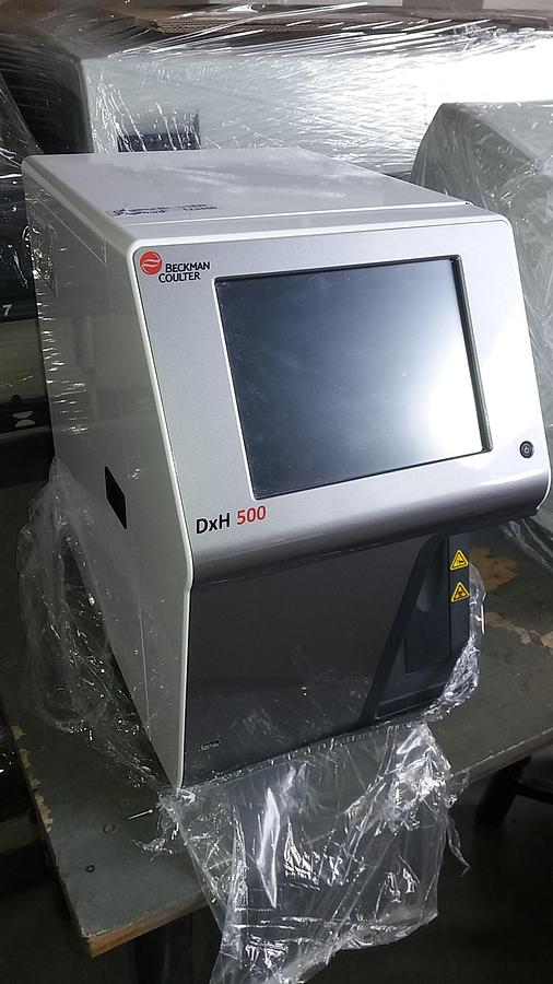
Beckman Coulter DXH 500
Details
Amishield veterinary chemistry analyzer
Details
For Sale FRESENIUS HEMOCARE 902407/F3009/COMPOMAT G 4
Details
For Sale HITACHI 704 Chemistry Analyzer
Details
BACTEC 9050
Details
SYSMEX KX 21
Details
ROCHE Cobas Integra 400 plus
Details
BIOMERIEUX Mini Vidas
Details
Viva-E
Details
Cormay, Accent 200
Details
ROCHE Connect 2 Plus
Details
ERBA XL-200
Details
SYSMEX CA-1500
Details
CORMAY Accent 200
Details
Sebia Capilarys 3 Tera
Details
Sebia Capilarys 3 Tera
Details
CORMAY Accent 200
Details
TOSOH HLC-723 G8
Details
Forfigate-50E FG-50E
Details
SIEMENS ADVIA CENTAUR CP
Details
For Sale BECKMAN COULTER PK 7300
Details
For Sale BECKMAN COULTER DxH 800 Hematology Analyzer
Details
Cobas c 702
Details
SYSMEX CA-1500
Details
Dade Behring, BEPIII
Details
For Sale DADE BEHRING BEP III
Details
Dade Behring, BEPIII
Details
For Sale SIEMENS BEP 2000
Details
Siemens, BEP2000
Details
TOSOH HLC-723G8
Details
Mediff hematological abacus
Details
Forfigate-50E
Details
COBAS INTEGRA 400 PLUS
Details
SIEMENS DCA Vantage
Details
TRINITY BIOTECH AMAX Destiny Plus
Details
SEBIA ASSIST
Details
BIOMERIEUX Vidas
Details
BIOMERIEUX Vidas Blue
Details
SIEMENS BNII
Details
SYSMEX SF-3000
Details
For Sale BACTEC 9240
Details
ABBOTT Alinity i
Details
2110 QiaGen QIA Cube
Details
MINDRAY BS-300
Details
Dako Omnis
Details
HORIBA pentra 60
Details
GEM Premier 4000
Details
ROCHE Cobas Mira Plus CC
Details
For Sale ROCHE Cobas Mira Plus CC Chemistry Analyzer
Details
CARL ZEISS Spekol 11
Details
For Sale CARL ZEISS Spekol 11 Unit
Details
SYSMEX OPSU-6
Details
SYSMEX OPSU-6
Details
AVL9120
Details
MPW-350R
Details
Xpress Piccolo
Details
For Sale CORNING CHLORIDE ANALYZER 925 Chemistry Analyzer
Details
For Sale SIEMENS BNII Chemistry Analyzer
Details
RADIOMETER ABL 800/805/810/815/820/825/837
Details
BAYER Rapidlab 348 EX
Details
OLYMPUS AU5421
Details
Cobas c513
Details
For Sale GRIFOLS DG THERM
Details
For Sale PACIFIC HEMOSTASIS ThromboScreen 400 C for in-vitro diagnostic
Details
TOSOH AIA-900
Details
For Sale HITACHI 7070 Chemistry Analyzer
Details
For Sale MINDRAY D-Chem 300 Chemistry Analyzer
Details
For Sale MAXMAT SA MAXMAT PL Chemistry Analyzer
Details
For Sale MINDRAY (Accent) BS 200 Chemistry Analyzer
Details
For Sale BECKMAN Paragon CZE 2000 Chemistry Analyzer
Details
For Sale MINDRAY BS-300 Chemistry Analyzer
Details
For Sale MINDRAY BS-300 Chemistry Analyzer
Details
For Sale BECKMAN COULTER UniCell DxC 600 Chemistry Analyzer
Details
BECKMAN COULTER UniCell DXC600
Details
For Sale ROCHE HITACHI 902 Chemistry Analyzer
Details
Viva-E
Details
For Sale SIEMENS VIVA E SYSTEM Chemistry Analyzer
Details
For Sale HORIBA Pentra 400 Chemistry Analyzer
Details
Dimension Xpand
Details
For Sale SIEMENS Dimension Xpand Chemistry Analyzer
Details
For Sale SIEMENS Dimension Xpand Plus Chemistry Analyzer
Details
For Sale SIEMENS ADVIA 1200 Chemistry Analyzer
Details
BIOSYSTEMS A15
Details
For Sale BIOSYSTEMS A15 Chemistry Analyzer
Details
For Sale BIOSYSTEMS A25 Chemistry Analyzer
Details
For Sale OLYMPUS AU 5800 Chemistry Analyzer
Details
For Sale SIEMENS Atellica CH 930 Chemistry Analyzer
Details
For Sale OLYMPUS Au400 Chemistry Analyzer
Details
For Sale ABAXIS VetScan Hematology Analyzer
Details
HPLC liquid chromatograph
Details
MB/BacT 120
Details
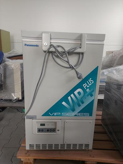
Panasonic MDF-C8V1 Low Temperature Freezer
Details
For Sale NIKON Eclipse 200 LED Microscope
Details
FOR SALE Siemens Clinitek Status
Details
FOR SALE Siemens Clinitek 500
Details
FOR SALE Siemens Clinitek Advantus
Details
FOR SALE Siemens Clinitek Atlas (Carousel)
Details
FOR SALE Maldi Tof MicroMass
Details
FOR SALE ABBOTT Cell-Dyn Ruby
Details
FOR SALE Siemens Rapidpoint 405
Details
FOR SALE Siemens Rapidpoint 350
Details
FOR SALE Siemens Rapidpoint 340
Details
FOR SALE Radiometer ABL 77
Details
FOR SALE Radiometer ABL 80 Flex
Details
For Sale BAYER Rapidlab/Ciba Corning 278/280 Blood Gas Analyzer
Details
For Sale SANYO MIR 162 Incubator
Details
FOR SALE SYSMEX KX 21N
Details
FOR SALE Siemens Rapidpoint 400
Details
FOR SALE Siemens DCA - 2000
Details
FOR SALE Siemens DCA 2000+
Details
FOR SALE SYSMEX KX 21
Details
FOR SALE Siemens Rapidchem744
Details
For Sale YUMIZEN H500 and H550 Hematology Analyzer
Details
For Sale BIOMERIEUX OPTION 2 Coagulation Analyzer
Details
Sysmex CellaVision XU-100027
Details
Hematology, Coulter T890
Details
For Sale CHATSWORTH ACP 100 USB Scanner
Details
For Sale BECKMAN SPINCHRON DLX - LAB Centrifuge
Details
SYSMEX CA-660
Details
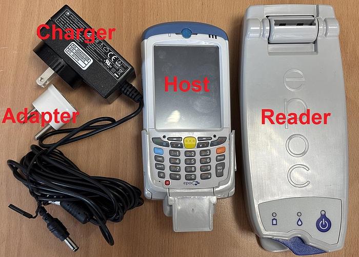
SIEMENS Epoc host Zebra MC55A0
Details
SIEMENS Immulite 1000
Details
DPC Immulite One
Details
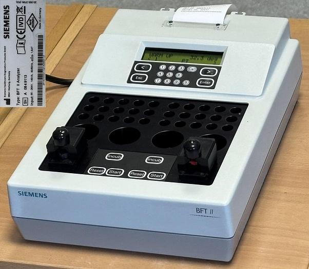
SIEMENS BFT II
Details
SIEMENS BFT II
Details
SIEMENS BFT II
Details
ROCHE DIAGNOSTICS MONITOR
Details
SIEMENS BFT II
Details
For Sale NELLCOR N-550 Oximeter - Pulse
Details
Erma EL-120 Electrolyte Analyzer
Details
ERMA INC PCE 210N
Details
Erma INCAUTO HEMATOLOGY ANALYZER PCE-370
Details
SYSMEX SP-1000I
Details
SIEMENS Immulite 2000
Details
SYSMEX pocH-100i
Details
SYSMEX pocH-100i
Details
SYSMEX pocH-100i
Details
TS-2000
Details
For Sale TREK DIAGNOSTIC SYSTEMS Sensititre AutoInoculator NEPHELOMETER
Details
DIASORIN LIAISON XL
Details
For Sale SIEMENS VersaCell X3 Solution
Details
For Sale SIEMENS BN PROSPEC Chemistry Analyzer
Details
For Sale BIOMERIEUX NucliSens EASYMAG
Details
For Sale BIOMERIEUX NucliSens Mini MAG
Details
For Sale SIEMENS STREAMLAB Full Automatic line for different analyzers
Details
For Sale BECKMAN COULTER OLA 2500 Chemistry Analyzer
Details
For Sale BECKMAN COULTER OLA 2500HS (High Speed) Chemistry Analyzer
Details
FOR SALE Olympus OLA 2500
Details
MUT HCTS2000
Details
For Sale ROCHE reflotron plus Chemistry Analyzer
Details
HUMAN Humalyzer 2000
Details
For Sale HUMAN - Humalyzer 3000 Chemistry Analyzer
Details
Incubator epoll 2
Details
Photometer 4040
Details
Kselmed 3002
Details
For Sale BIOMERIEUX Mini Vidas Barcode Reader/ Scanner
Details
For Sale BODITECH MED i-CHROMA Reader Blood Analyzer
Details
Boditech I Chroma
Details
For Sale HUMAN 315 Humareader Single
Details
For Sale BERTHOLD TECHNOLOGIES The Lumat LB 9507 tube luminometer
Details
For Sale TOSOH HLC-723G8 Hemoglobinometer
Details
For Sale SHORELINE SMP41 Refrigerator Freezer
Details
For Sale ACCUSCIENCE Blood Track Kiosk V4
Details
For Sale JEWETT BBR37 Refrigerator Freezer
Details
Thermo Electron Corporation Incubator Hepa Class 100
Details
For Sale MINDRAY BC-3000 Hematology Analyzer
Details
For Sale BECKMAN COULTER LH-750 Hematology Analyzer
Details
For Sale BECKMAN COULTER LH 500 Hematology Analyzer
Details
For Sale BECKMAN COULTER STKS Hematology Analyzer
Details
For Sale BECKMAN COULTER ACT 5 Diff AL Hematology Analyzer
Details
For Sale BECKMAN COULTER ACT 5 Diff CP Hematology Analyzer
Details
For Sale BECKMAN COULTER ACT 5 diff Hematology Analyzer
Details
For Sale HORIBA Micros 60ES - opened system Hematology Analyzer
Details
For Sale ABX Pentra 120. Hematology Analyzer
Details
For Sale HORIBA pentra 60 Hematology Analyzer
Details
For Sale COULTER T-660 Hematology Analyzer
Details
For Sale COULTER T-890 Counter Hematology Analyzer
Details
For Sale COULTER Micro Diff II Hematology Analyzer
Details
For Sale MEDONIC CA-530 Boule Medical Hematology Analyzer
Details
For Sale MEDONIC CA 530-16 Hematology Analyzer
Details
FOR SALE ABBOTT Cell-Dyn Sapphire
Details
For Sale CELL DYN Sapphire Hematology Analyzer
Details
SYSMEX K-4500
Details
For Sale SYSMEX K-4500 Hematology Analyzer
Details
For Sale SYMBOL LS2208 General Purpose Barcode Scanner
Details
For Sale BINDER Dryer
Details
For Sale COBAS EIA Incubator
Details
AFS8D WATER PURFICATION
Details
Millipore ELIX 3 Station Water
Details
For Sale MILLIPORE Elix 10
Details
For Sale MILLIPORE ELIX 10
Details
For Sale MILLIPORE ELIX 20
Details
For Sale MILLIPORE AFS-30
Details
For Sale MILLIPORE AFS 10D
Details
For Sale MILLIPORE AFS 16D
Details
For Sale MILLIPORE AFS 8D
Details
For Sale COBAS 6000 Chemistry Analyzer
Details
For Sale KODAK Ektachem DTSC II MODULE Chemistry Analyzer
Details
For Sale BAYER DCA 2000+ Chemistry Analyzer
Details
For Sale REFLOTRON IV / PLUS Chemistry Analyzer
Details
For Sale BIO-RAD PR 4100 Absorbance Microplate Reader
Details
MINDRAY MW-12A
Details
For Sale MINDRAY 12A Minrday 12A Microplate washer
Details
For Sale HEIDOLPH TITRAMAX 1000 Platform Shaker Rotator/Mixer/Rocker
Details
For Sale DIAMOND DIAGNOSTICS SMARTLYTE Electrolyte Analyzer
Details
For Sale ROCHE 9180 Electrolyte Analyzer
Details
For Sale CORNING 480 Electrolyte Analyzer
Details
For Sale AVL 9110 Electrolyte Analyzer
Details
For Sale AVL 9180 Electrolyte Analyzer
Details
For Sale IEC MULTI-RF Centrifuge
Details
For Sale ROCHE LC Carousel Centrifuge
Details
For Sale BAXTER Immufuge II Centrifuge
Details
For Sale EPPENDORF 5403 Centrifuge
Details
For Sale VANGUARD V-6500 Centrifuge
Details
For Sale SORVALL MIC-0019E Legend Centrifuge
Details
For Sale MTS MPW 340 Centrifuge
Details
For Sale JOUAN CR-412 Centrifuge
Details
For Sale JOUAN C-412 Centrifuge
Details
Diamed Centurfige 24S
Details
For Sale SIEMENS Immulite 1000
Details
For Sale BIOMERIEUX Mini Vidas Blue/Grey
Details
DIASORIN Liaison
Details
For Sale DIASORIN Liaison YOM 2011
Details
Immulite 1000
Details
For Sale ABBOTT LABS Architecti2000
Details
For Sale SIEMENS ADVIA Centaur XP Immunology Analyzer
Details
For Sale BAYER ADVIA CENTAUR - XP/CP
Details
For Sale SIEMENS Centaur Classic Immunology Analyzer
Details
For Sale SIEMENS ADVIA CENTAUR CP
Details
For Sale ABBOTT IMX SYSTEM
Details
For Sale PHADIA ImmunoCAP 250
Details
For Sale ROCHE Cobas E601
Details
Siemens Atellica IM 1600
Details
For Sale BECKMAN COULTER UniCel DXI 800
Details
For Sale SIEMENS Atellica Direct Load Chemistry Analyzer
Details
For Sale SIEMENS Atellica IM 1300 Analyzer & Atellica IM 1600 Chemistry Analyzer
Details
For Sale POINTE 180 Chemistry Analyzer
Details
For Sale URISYS 1800 Urine Analyzer
Details
For Sale SIEMENS STATUS PLUS Urine Analyzer
Details
For Sale SIEMENS Clinitek STATUS + Urine Analyzer
Details
For Sale BAYER clinitek Status plus Urine Analyzer
Details
For Sale ROCHE Urisys 2400 Urine Analyzer
Details
For Sale BAYER clinitek advantus Urine Analyzer
Details
Clinitek Advantus
Details
For Sale DIGITAL Diagnostic Stago STart4
Details
For Sale INSTRUMENTATION LABORATORY ACL ELITE/ELITE PRO Coagulation Analyzer
Details
For Sale SYSMEX CA-550 Coagulation Analyzer
Details
For Sale INSTRUMENTATION LABORATORY ACL 7000 Coagulation Analyzer
Details
For Sale INSTRUMENTATION LABORATORY ACL 7000 Coagulation Analyzer
Details
For Sale BECKMAN COULTER ACL ADVANCE Coagulation Analyzer
Details
For Sale INSTRUMENTATION LABORATORY ACL 10000 Coagulation Analyzer
Details
For Sale INSTRUMENTATION LABORATORY ACL 8000 Coagulation Analyzer
Details
For Sale SYSMEX CA-540 Coagulation Analyzer
Details
FOR SALE Dade Behring BCT
Details
For Sale DADE BEHRING BCT Coagulation Analyzer
Details
FOR SALE Sysmex CA 560
Details
For Sale SYSMEX CA-560 Coagulation Analyzer
Details
CS 5100 SYSMEX Coagulology Analyzer
Details
For Sale BAXTER Walk Away-40 Micro S
Details
For Sale BAXTER Walk Away 96 Microscan
Details
For Sale BIOMERIEUX VITEK 2 COMPACT
Details
SIEMENS BCS XP
Details
For Sale VITALOGRAPH Compact Spirometer
Details
For Sale JAEGER Spirometr Flowscreen Spirometer
Details
For Sale VIASYS Flowscreen II Spirometer
Details
For Sale PARI TURBO BOY Respirator
Details
SIEMENS BCS XP
Details
SIEMENS BCS XP
Details
For Sale BATH CHAIR Ferno ille 100 Bath Chair
Details
For Sale THERMO SCIENTIFIC FORMA 88000 SERIES Refrigerator Freezer
Details
For Sale RADIOMETER OSM-3 Blood Gas Analyzer
Details
For Sale ROCHE OMNI C/ Cobas b121 Blood Gas Analyzer
Details
For Sale AVL OMNI Blood Gas Analyzer
Details
For Sale RADIOMETER EML 105 Blood Gas Analyzer
Details
For Sale RADIOMETER ABL-510 Blood Gas Analyzer
Details
For Sale RADIOMETER ABL 330 Blood Gas Analyzer
Details
For Sale RADIOMETER ABL 800 Blood Gas Analyzer
Details
For Sale SIEMENS Rapidpoint 405 Blood Analyzer
Details
For Sale BAYER Rapidpoint 400/405 Blood Gas Analyzer
Details
Elkon Cl-65
Details
For Sale SYSMEX UF 1000i Urine Analyzer
Details
For Sale SYSMEX UF 100 Urine Analyzer
Details
For Sale BARD Sherlock Vascular - Small Parts Ultrasound
Details
For Sale ALOKA ECHO CAMERA SSD-210 DX
Details
For Sale GRIFOLS Wadiana .
Details
For Sale GRIFOLS Erytra
Details
For Sale ROCHE Cobas Mira S Chemistry Analyzer
Details
For Sale DADE BEHRING Stratus CS. Chemistry Analyzer
Details
For Sale HITACHI 705 Chemistry Analyzer
Details
For Sale VITROS 950 AT Chemistry Analyzer
Details
For Sale SIEMENS-ELEMA CA 930 Co2 Analyzer
Details
For Sale HANNA ELECTRODE HOLDER
Details
Amelung CS 190 Amax
Details
AUTO HEMATOLOGY ANALYZER PCE-210N
Details
For Sale SYSMEX KX-21N Hematology Analyzer
Details
OPTA-TECH N-180
Details
For Sale ALOKA 4000
Details
MPW-223e
Details
HORIBA ABX ABX Micros 60 OT
Details
Rotanda 460R
Details
ThermoFisher Brah Kryptor
Details
Abbott TDX
Details
Architect i1000SR
Details
Magna Pure ROCHE DIAGNOSTICS GmbH JE379
Details
PZO WARSZAWA Microscope
Details
PZO WARSZAWA Microscope
Details
Magisculpt Manual Sm-20
Details
UPS Sinergy 2 SE04XH
Details
SIEMENS Rapidlab 348 EX
Details
SEBIA Hydrasys
Details
Sebia Hydrasys
Details
Thermo Scientific™ Orion™ ROSS Ultra™ Glass Bodied Combination pH Electrode
Details
Thermo Orion 081010MD Ross combination pH electrode
Details
For Sale THERMO ELECTRON RUSSEL RL060P Portable pH Meter PH Meter
Details
THERMO ELECTRON RUSSEL RL060P Portable pH Meter
Details
Orion 3 Star Portable Ph meter
Details
Orion Orion Star A326
Details
Orion 4 Star Benchtop pH/ISE meter
Details
ORION 3 Star Benchtop pH meter
Details
For Sale ORION 5-STAR pH/ORP/ISE/Conductivity Electrolyte Analyzer
Details
For Sale ORION 3-Star series Electrolyte Analyzer
Details
For Sale PWBHEALTHLTD Breastlight breast awareness
Details
BreastLight a Device for performing Breast Examination Using Light
Details
Pari TurboBoy Specialists in Effective Inhalation
Details
Feya Device for Thermotherapy of Accessory Nose and Laryngeal Sinuses
Details
UTMpk-01 Device for Rectum Thermomagnetic Therapy
Details
Almag-01 Portable Magnetotherapy Device with a Travelling Pulsed Magnetic field
Details
For Sale SYSMEX UF-100 Urine Analyzer
Details
For Sale SYSMEX UF-50 Urine Analyzer
Details
SYSMEX UF500i
Details
SIEMENS Dimension EXL 200
Details
Dimension RXL MAX
Details
Sysmex Expertline HST-N310
Details
Sebia Hydrasys
Details
For Sale SEBIA Hydrasys
Details
Sebia Hydrasys
Details
SEBIA Hydrasys
Details
Hydrasys
Details
Sebia Hydrasys
Details
For Sale IL ILAB 300 Plus Chemistry Analyzer
Details
For Sale COBAS INTEGRA 800 Chemistry Analyzer
Details
For Sale SIEMENS CLINITEK NOVUS Chemistry Analyzer
Details
Siemens Atellica 1500 Automated Urinalysis System
Details
For Sale DPC VersaCell SMS Chemistry Analyzer
Details
For Sale OLYMPUS AU 600 Chemistry Analyzer
Details
For Sale VITROS 250 Chemistry Analyzer
Details
For Sale TECHNICON RA 1000 Chemistry Analyzer
Details
For Sale METROLAB 2300 PLUS Chemistry Analyzer
Details
For Sale BIO-RAD D Chemistry Analyzer
Details
For Sale VITALAB FLEXOR E Chemistry Analyzer
Details
For Sale VITAL DIAGNOSTICS FLEXOR E Chemistry Analyzer
Details
Zeenit ZeemanAAS Spectrometer-Jena
Details
For Sale THERMO FINNIGAN TSQ Quantum Ultra Spectrophotometer
Details
For Sale ELAMED FEYA Device Thermotherapy of Accessory Nose
Details
For Sale ELAMED TEPLON device for Thermotherapy
Details
For Sale THERMO SCIENTIFIC 40 AFT Lab-Tower
Details
For Sale BIOMERIEUX Konelab 20i Chemistry Analyzer
Details
For Sale THERMO KONELAB Konelab 30i Chemistry Analyzer
Details
THERMO SCIENTIFIC Konelab 30i
Details
BIOMERIEUX Vidas
Details
For Sale COBAS EIA Spectrophotometer
Details
For Sale EPPENDORF PCP 6121 Spectrophotometer
Details
For Sale ANALYTIK JENA AG LP 300 DR Lange. Spectrophotometer
Details
For Sale THERMO SCIENTIFIC MULTISKAN EX Spectrophotometer
Details
For Sale APTUS 466
Details
For Sale WATERS INSTRUMENTS MICROMASS MALDI TOF Spectrophotometer
Details
Cobas Integra 400+
Details
For Sale BIO-RAD Evolis 6370
Details
MINDRAY MR - 96A
Details
SIEMENS Epoc Reader
Details
Epoc Host
Details
For Sale CORNING CIBA 604 AUTOSAMPLER
Details
For Sale GRANT JB Aqua 12 Water Bath
Details
For Sale MELAG THERMOSTAT LEIHGERAET 71
Details
Sensititre Autolnoculator
Details
For Sale TREK DIAGNOSTIC SYSTEMS Sensititre Sensitouch Autoinoculator
Details
For Sale MOLECULAR DEVICES DPC MILENIA KINETIC ANALYZER Microplate Reader
Details
For Sale TECAN Spectra Mini AP Microtiter plate photometer
Details
For Sale DYNEX DSX Microplate Reader
Details
ELGA USF MEDICA 30
Details
For Sale SIEMENS Hematek 2000 Slide Stainer
1
2
›
»
To the top